A clear focus on the problem
Cone beam–computed tomography overcomes the limitations of conventional radiographs for endodontics, writes Brian Vaughan
The use of cone beam–computed tomography (CBCT) is rapidly increasing in dentistry to aid in diagnosis and treatment planning for a variety of clinical situations. Implants, endodontics, oral surgery, ortho–dontics, periodontics, treatment of TMD or treatment of sleep apnoea are the m
ain specialties in which CBCT is currently used1.
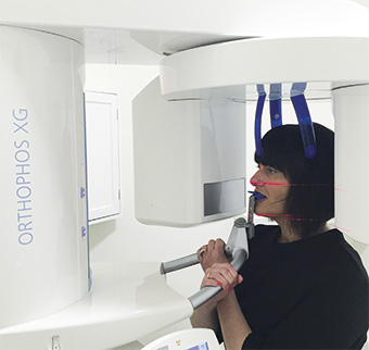
At Boyne Dental and Implant Clinic, as part of the relocation to new premises in October 2015, a Sirona XG 3D CBCT scanner was purchased. This new technology has already proved to be invaluable in the practice for the treatment of implant and endodontic patients as will be shown in a recently treated case.
Limitations of radiographs
Intra–oral radiographs remain the standard imaging modality for diagnosis, managing and assessing endodontic disease2. However, radiographs only provide a 2D image of a 3D structure and therefore have several limitations. These include:
- compression of anatomy3 as the image is only viewed in a mesio–distal (proximal) plane and the bucco–lingual/palatal plane is not observed
- anatomical noise4 produced by overlying structures such as zygomatic buttress or cortical bone
- geometric distortion4, for example, elongation of the image due to a shallow palatal vault
- temporal perspective as the positioning of intra–oral radiographs cannot be standardised a slight change in angulation can give a false sign that there is radiographic healing of an apical lesion5.
Applications in endodontics
CBCT overcomes the problems associated with conventional radiographs. It is always desirable that the field of view (FOV) should be as small as possible and limited to the area of interest thus reducing the radiation exposure to the patient7. In endodontics this is usually 5x5cm and the use of a limited volume CBCT scanner, e.g. Sirona XG 3D, makes this achievable. As shown in Table 1, the radiation dose for a 5×5.5cm scan is comparable to between four and eight periapical radiographs.
The images produced from the CBCT are a true 3D representation of the area of interest. The lower radiation dose compared to traditional CT scanners8 and geometric accuracy of scanned objects 9,10 makes CBCT ideal for treatment planning apical surgery11, diagnosis of dento–alveolar trauma and in certain cases the diagnosis of radiolucent apical lesions12.
The European Society of Endodontology position statement on the use of CBCT in Endodontics13 describes the following indications:
- Diagnosis of radiographic signs of periapical pathosis when there are contradictory (non–specific) signs and/or symptoms
- Confirmation of nonodontogenic causes of pathosis
- Assessment and/or management of complex dento–alveolar trauma
- Appreciation of extremely complex root canal systems prior to endodontic management e.g. Dens Invaginatus (Case 1)
- Assessment of endodontic treatment complications e.g. post perforations, for treatment planning purposes
- Assessment/management of root resorption which clinically appears to be amenable to treatment (Cases 2 and 3)
- Pre–surgical assessment prior to complex periradicular surgery.
For information on the current European guidelines for referring dentists and for operators of CBCT, please visit www.sedentexct.eu
Case report
A 54–year–old female patient was referred complaining of pain and tenderness from her lower left first premolar (34). Root canal treatment had been completed three weeks previously. On examination there was acute tenderness to percussion, negative sensibility testing and no buccal or lingual swelling. Radiographically there was a well obturated canal of good taper and length. There was slight asymmetry of the root filling and this can be indicative of a second canal.
On close inspection of the radiograph, a second canal could be detected. Rubber dam had been used and the canal prepared with ProTaper rotary files and irrigated with sodium hypochlorite. Obturation was with gutta percha and AH Plus (Dentsply) using a cold lateral condensation technique.
Mandibular premolar teeth exhibit a great variation in both root canal and root morphology compared to the conventional one root one canal found in Dental Anatomy textbooks14.
Vertucci (1984) 15 found that in 400 mandibular first premolars investigated 74 per cent had one canal at the apex, 25.5 per cent had two canals and 0.5 per cent three canals. The incidence of two separate canals (Type IV configuration, see Fig 1) was 1.5 per cent.
One of the main reasons for endodontic failure is inadequate instrumentation and cleaning of the root canal system16 and, due to the anatomical variations, the lower mandibular premolar has been described as presenting one of greatest the challenges to successfully treat17.
A 5×5.5cm CBCT scan (Sirona XG 3D) was taken to confirm the presence and location of the second canal. The root canals tend to be behind superimposed on a radiograph as they are behind each other in the bucco–lingual plane18. The CBCT images showed there was a second root and canal located lingually (Fig 2). Under rubber dam and using microscope magnification the lingual canal was identified (Fig 3a). The canal was prepared with K files and rotary Wave One Gold Primary file (Fig 3b).
The irrigation protocol was copious sodium hypochlorite activated sonically with Endoactivator (Dentsply) and a penultimate rinse with 17 per cent EDTA solution19. The Master Apical File size was 40. Two weeks later, after dressing with calcium hydroxide and glass ionomer, the symptoms had resolved. Under rubber dam the canal was accessed, working length and patency checked and irrigated with the above protocol.
The canal was dried with sterile paper points (Dentsply) and obturated with gutta percha and AH Plus sealer (Dentsply) using the warm vertical compaction technique (B&L Biotech) (Fig 3c). The access cavity was restored with SDR (Dentsply) and Venus Diamond (Heraeus Kulzer). A post–operative radiograph was taken and two separate canals are satisfactorily obturated. The patient has been advised that a cuspal coverage restoration is required to provide the best long–term prognosis20,21.
Summary
The integration of CBCT into Boyne Dental and Implant Clinic has increased the predictability of the treatment carried out. By providing a more accurate diagnosis there is added confidence that a successful treatment outcome will be achieved. It has already proved to be an extremely useful addition to the armamentarium for the management of complex endodontic problems.
About the author
Brian Vaughan qualified from Queen’s University Belfast. After working in England for three years he returned to Northern Ireland where he worked as a senior house officer in restorative dentistry and a special lecturer in the school of dentistry, and then in private practice.
Brian is a dentist with special interest in endodontics and he is currently studying a Masters degree in Endodontics at King’s College London. Since 2010 he has been using a dental operating microscope and taking referrals for endodontic treatment. His practice is now limited to endodontics in Boyne Dental and Implant Clinic, Navan. He was recently one of the speakers at Sirona Innovations Day in Henry Schein Dublin on CBCT and endodontics.
References
1. Cotton TP, Geisler TM, Holden DT, Schwartz SA, Schindler WG. Endodontic applications of cone–beam volumetric tomography. Journal of Endodontics. 2007 Sep;33(9):1121–32.
2. Lofthag–Hansen S, Huumonen S, Gröndahl K, Gröndahl H–G. Limited cone–beam CT and intra–oral radiography for the diagnosis of periapical pathology. Oral Surgery, Oral Medicine, Oral Pathology, Oral Radiology, and Endodontology. 2007 Jan;103(1):114–9.
3. Patel S, Dawood A, Mannocci F, Wilson R, Pitt Ford T. Detection of periapical bone defects in human jaws using cone beam computed tomography and intra–oral radiography. Int Endod J. 2009 Jun;42(6):507–15.
4. Gröndahl H–G, Huumonen S. Radiographic manifestations of periapical inflammatory lesions. Endodontic Topics. Munksgaard International Publishers; 2004 Jul 1;8(1):55–67.
5. Rudolph DJ, White SC. Film–holding instruments for intra–oral subtraction radiography. Oral Surg Oral Med Oral Pathol. 1988 Jun;65(6):767–72.
6. Ludlow JB, Timothy R, Walker C, Hunter R, Benavides E, Samuelson DB, et al. Effective dose of dental CBCT– a meta analysis of published data and additional data for nine CBCT units. Dentomaxillofacial Radiology. 2015 Jan;44(1):20140197–25.
7. Palomo JM, Rao PS, Hans MG. Influence of CBCT exposure conditions on radiation dose. Oral Surgery, Oral Medicine, Oral Pathology, Oral Radiology, and Endodontology. Elsevier; 2008 Jun;105(6):773–82.
8. Mozzo P, Procacci C, Tacconi A, Martini PT, Andreis IAB. A new volumetric CT machine for dental imaging based on the cone–beam technique: preliminary results. European Radiology. Springer–Verlag; 1998;8(9):1558–64.
9. Mischkowski RA, Pulsfort R, Ritter L, Neugebauer J, Brochhagen HG, Keeve E, et al. Geometric accuracy of a newly developed cone–beam device for maxillofacial imaging. Oral Surgery, Oral Medicine, Oral Pathology, Oral Radiology, and Endodontology. 2007 Oct;104(4):551–9.
10. Eggers G, Klein J, Welzel T, Mühling J. Geometric accuracy of digital volume tomography and conventional computed tomography. Br J Oral Maxillofac Surg. Elsevier; 2008 Dec;46(8):639–44.
11. Patel S, Dawood A. The use of cone beam computed tomography in the management of external cervical resorption lesions. Int Endod J. Blackwell Publishing Ltd; 2007 Sep;40(9):730–7.
12. Simon JHS, Enciso R, Malfaz J–M, Roges R, Bailey–Perry M, Patel A. Differential Diagnosis of Large Periapical Lesions Using Cone–Beam Computed Tomography Measurements and Biopsy. Journal of Endodontics. 2006 Sep;32(9):833–7.
13. Patel S, Durack C, Abella F, Roig M, Shemesh H, Lambrechts P, et al. European Society of Endodontology position statement: The use of CBCT in Endodontics. Int Endod J. 2014 Jun 1;47(6):502–4.
14. Cleghorn BM, Christie WH, Dong CCS. Anomalous mandibular premolars: a mandibular first premolar with three roots and a mandibular second premolar with a C–shaped canal system. Int Endod J. 2008 Nov;41(11):1005–14.
15. Vertucci FJ. Root canal anatomy of the human permanent teeth. Oral Surgery, Oral Medicine, Oral Pathology. 1984;58(5):589–99.
16. Chan K, Yew SC, Chao SY. Mandibular premolar with three root canals – two case reports. Int Endod J. 1992 Sep;25(5):261–4.
17. Lu T–Y, Yang S–F, Pai S–F. Complicated Root Canal Morphology of Mandibular First Premolar in a Chinese Population Using the Cross Section Method. Journal of Endodontics. 2006 Oct;32(10):932–6.
18. Robinson S, Czerny C, Gahleitner A, Bernhart T, Kainberger FM. Dental CT evaluation of mandibular first premolar root configurations and canal variations. Oral Surgery, Oral Medicine, Oral Pathology, Oral Radiology, and Endodontology. 2002 Mar;93(3):328–32.
19. Haapasalo M, Shen Y, Qian W, Gao Y. Irrigation in endodontics. Dental Clinics of North America. 2010 Apr;54(2):291–312.
20. Sorensen JA, Martinoff JT. Intracoronal reinforcement and coronal coverage: a study of endodontically treated teeth. J Prosthet Dent. 1984 Jun;51(6):780–4.
21. Salehrabi R, Rotstein I. Endodontic Treatment Outcomes in a Large Patient Population in the USA: An Epidemiological Study. Journal of Endodontics. 2004 Dec;30(12):846–50.

