Involving the patient in the process
Stuart Campbell BDS, MFDS RCS (Edin), describes how he used a modified approach to replace a patient’s missing maxillary incisor
The replacement of a missing maxillary central incisor in a patient with diastemata can be problematic for the restoring dentist. To achieve a successful outcome, a modified approach to treatment may be necessary.
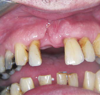
This case report describes a 53-year-old male with chronic periodontal disease, a moderate gag reflex and diastemata who presented in general practice with a missing upper right central incisor, 11. This tooth had exfoliated secondary to a combined periodontal and endodontic lesion (Fig 1). An unsuccessful attempt had been made to treat the lesion with endodontic and periodontal therapy (Fig 2).
The patient had brought the exfoliated tooth with him which revealed an intact exfoliation.
The patient’s medical history was unremarkable. The smile line revealed the entire surface of the upper incisors and around 1mm of attached gingivae. Intra-oral examination revealed a moderate gag reflex which could be triggered during routine examination.
Further examination revealed a moderately restored dentition with multiple spacing or diastemata. The dentition was intact with the exception of the recently exfoliated upper right central incisor. The patient had chronic generalised periodontitis with moderate to severe bone loss. There was moderate loss of tissue volume secondary to periodontal disease and exfoliation of 11.
There was right group function and left canine guidance with protrusive contacts noted on the upper and lower incisors. No posterior protrusive interferences were noted. The teeth adjacent to the missing incisor were unrestored, exhibited Grade I+ mobility1 and responded positively to vitality tests. The patient requested that his diastemata were preserved.
The treatment options for the replacement of the missing central incisor are well documented and are as follows:
- do nothing
- dental implant
- removable prosthesis
- fixed conventional bridge
- fixed adhesive bridge.
Do nothing
In order to obtain informed consent, the option of no treatment should always be offered to the patient3. In this case, the patient declined to have no treatment and requested the missing tooth was restored.
Dental implant
The well-documented success rates of dental implants4 has led to these restorations being described as the most effective replacements for single missing incisors5,8.
However, dental implants are not suitable for all patients and may be contraindicated due to cost, smoking, systemic health problems such as osteogenesis imperfecta, bisphosphonate therapy, radiotherapy and severe diabetes6. In this case the use of dental implants would be ill-advised due to the presence of active periodontal disease, which can negatively affect osseointegration7.
Removable appliance
A removable appliance is a cheap, relatively straightforward way to replace a missing tooth and can provide excellent aesthetics8. However, in this case such an appliance was inappropriate due to the patient’s gag reflex.
Fixed bridgework
The successful use of fixed adhesive and conventional bridgework to restore patients with compromised periodontal support is well documented9,10.
The problem in this case was the size of the edentulous space. Neither the options of a very wide pontic or the re-distribution of space through conventional preparation of all of the maxillary incisors or with orthodontics was considered desirable.
Planning
After discussing the options it was clear that the patient favoured the use of minimal preparation adhesive bridgework to restore the space.
A conventional bridge design was inappropriate because of the patient’s desire to maintain his diastema. With the conventional design, the pontic and retainer are soldered together, leaving no space for preservation of diastema. This issue was addressed by planning a modified spring cantilever adhesive bridge as described by Gibson11.
It was considered appropriate to keep the length of the spring cantilever arm short, to avoid triggering the gag reflex and to increase the rigidity of the prostheses. For these reasons, together with aim of being minimally invasive, a conventional spring cantilever bridge was dismissed.
Treatment
Impressions were taken for study casts. The use of modified stock trays and distraction techniques were used to manage the patient’s gag reflex12,13. Study casts were articulated with the aid of a facebow registration and the technician was instructed to perform a diagnostic wax up.
A fixed/fixed bridge design was proposed on units 12, 21 and shown to the patient with the aid of the diagnostic wax up. This design was considered to minimise rotational forces on the pontic during protrusion and prevent future drifting of the abutments2. Pink porcelain was planned to replace the lost tissue volume which was visible during smiling. The wax-up revealed the need to further augment the pontic with pink porcelain to ensure symmetry (Fig 3).
An alternative design may have been to use tooth 13 as the retainer, however the patient considered the splinting effect of the fixed/fixed design to be advantageous. After patient approval of the bridge design, operative treatment was planned. Minimal preparation of the palatal surfaces of 12 and 21 was carried out using the speed increasing handpiece without local anaesthetic.
The preparation involved supragingival chamfer margins and parallel slots to increase resistance and retention form. Definitive Impressions were taken in modified stock trays using Polyether. The gag reflex was again managed with distraction techniques.
The technician was instructed to make a three-unit fixed/fixed spring cantilever adhesive bridge with pink porcelain to restore lost tissue (Fig 4, 5). The shade, characteristics and texture of porcelain for the pontic were prescribed. The shade of pink porcelain was chosen using a customised shade guide supplied by the dental technician. At the patient’s request no provisional restoration was provided.
Following laboratory manufacture, try-in of the appliance was carried out and an accurate fit of the framework was observed. Occlusal excursions were checked and adjustments were made to minimise protrusive contacts on the pontic (Fig 6).
Following try-in, the fit surface of the bridge was sandblasted using 50ʯm alumina in a chairside air abrasion system (Prophy-mate Neo NSK).
The use of rubber dam is considered essential for adhesive bridge cementation (14) A three-dimensional rubber dam system with anatomical frame was applied using the split dam technique (Fig 7, 8).
Once isolation was achieved, the abutment teeth were cleaned with prophylaxis paste. The bridge was cemented using adhesive cement and excess cement was removed. Metal margins were burnished to the tooth structure using fine diamond burs in the air-rotor. The use of copious water spray during finishing procedures is recommended to avoid the generation of excessive heat which can negatively affect the strength of the composite bond to both tooth and metal14. Oral hygiene instruction was provided and review appointments were arranged. The patient was satisfied with the final result (Figs 9, 10, 11, 12).
Discussion
Replacing a missing maxillary anterior tooth in a spaced dentition is a challenge for the restoring dentist. This case illustrates the importance of giving consideration to all treatment options while involving the patient in the decision making process.
The use of the adhesive spring cantilever bridge provided this challenging patient with a satisfactory outcome while preserving tooth tissue. This design may be considered by restoring dentists when faced with similar cases in practice or as a provisional appliance prior to implant restoration.
Acknowledgements:
All clinical dentistry was carried out by Mr Stuart Campbell, Loanhead Dental Practice,Midlothian. Laboratory work carried out by Mr Kenneth Scott, A-Plus Dental Laboratory, Dundee.
About the author
Stuart Campbell qualified from University of Dundee in 2001 and is a vocational trainer and partner at Loanhead Dental Practice. He is currently studying for an MSc in Implant Dentistry at the University of Central Lancashire.
To see all the clinical photographs from this article, visit out Facebook site by clicking here.