The science of a dazzling smile
In the second part of his series, Dr Paul A. Tipton looks at the problems associated with failing post crowns
In the previous article in this series, we looked extensively at the problems associated with replacing a post crown with an implant as an immediate replacement and restoration in the anterior maxilla. There are numerous other problems when confronted by a failing post crown with infection that needs to be treated in a different manner: that of removal, waiting for healing of up to three months, followed by implant placement and restoration some two to four months later. This article will highlight some of these problems by looking at a typical case study.
Case presentation
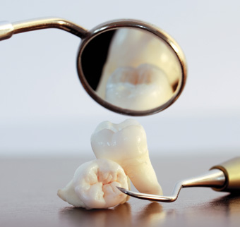
Patient A (Fig 1) was referred to my dental clinic by her general dental practitioner. Her complaint was that she had infection around a post crown in the upper right central incisor position (Fig 2), and that she was unhappy with her gummy smile and irregular-looking upper anterior teeth (Fig 3).
A full occlusal examination revealed good posterior teeth and stability with no signs of TMJ/bruxing/wear. So the decision was made to work in a conformative rather than a reorganised manner around the patient’s existing intercuspal position (ICP).
The post crown and infected tooth root were removed and the bone and soft tissues were left to heal for three months while the patient wore a simple partial denture. After this time, crown-lengthening procedures were completed to eliminate the gummy smile using the position of the gingival margins of the canine teeth as a reference. At the same time as the crown lengthening, an implant was placed into the upper right central inner region.
Shortly after crown lengthening (10 days), the teeth were prepared and prototype-linked composite crowns were placed on the five remaining upper anterior teeth and the implant healing abutment (Fig 4). These prototypes were in place for a further three months while the soft tissue healed after surgery, when further impressions were taken for a Procera Zirconia abutment for the implant.
After a further month for maturation of the soft tissues, further definitive tooth preparations were completed on the remaining upper anterior teeth for the construction of dentine-bonded feldspatic porcelain crowns. This was immediately followed by hybridisation (bonding) of the dentine to seal all tubules. Removal of the oxygen inhibition layer was completed by further curing of the dentine bond after placement of glycerine on to the preps. This step is essential so that the impression material will set completely.
The implant abutment was restored with a Procera alumina crown and the other five teeth had feldspatic porcelain 360 degree veneers (dentine bonded crowns) fitted with composite cuting cement.
The final result shows an even appearance of the crowns with the elimination of the gummy smile and replacement of the infected post crown by an implant (Figs 5, 6, 7).
Conclusions
In a case like this where the infected post crown is removed and left to heal, there are many potential aesthetic problems. These include the final positioning of the interdental peaks of bone whose blood supply comes from, in part, the periodontal ligament. When the tooth is removed, these precious pieces of bone and associated length of interdental papillae are lost.
Thus the delayed implant placement restoration often has to have long contact points to overcome the lack of interdental papillae.
Other problems include vertical and horizontal bone loss due to infection, which will compromise the vertical and horizontal placement of the implant. In a case like this one, where fortunately there is a gummy smile that requires soft and hard tissue excision, the problem of vertical bone loss is overcome.
Should a gummy smile not be present then the use of extensive soft tissue reconstructive surgery and/or pink porcelain may be required. The next article in the series will deal with these solutions.
Finally, horizontal bone loss will mean the implant is placed further to the palatal with problems of emergence profile in order to get the tooth into the correct position.
Again, soft tissue surgery may be required to build up the ridge, followed by excision to allow better emergence profile. Cases where there are horizontal defects will also be discussed in the next in this series of articles.
ABOUT THE AUTHOR
A highly respected specialist in prosthodontics, Dr Paul Tipton has published many scientific articles in the dental press and is an expert lecturer in his field with Tipton Training Academies in Dublin, Manchester, Leeds and London. After gaining his masters degree in conservative dentistry in 1989, he was awarded the diploma in general dental practice by the Royal College of Surgeons four years later and received specialist status in prosthodontics in 1999 from the GDC. An ex-professional cricketer with Lancashire County Cricket Club, he is a past president of the British Academy of Implant Dentistry (http://www.baid.org.uk).
Dr Tipton is one of the UK’s most successful dental teachers in the fields of restorative, cosmetic and implant dentistry over the past 20 years with more than 2,000 dentists completing year long certificate courses from one of the Tipton Training Academies (tiptontraining.co.uk).
Dr Tipton would like to thank the following people for help with this series of articles: • Dr Ibrahim Hussain, BDS, M Med Sci Implantology – implant surgeon
• Dr Andrew Watson, BDS, MSc, specialist in endodontics
• Mr Jeff Caddick, dental technician, Castle Ceramics, Staffordshire
• Dr Amit Patel, BDS, MSc, MClinDent, MFDS, RCSEd , MRD, RCSEng specialist in periodontics.