
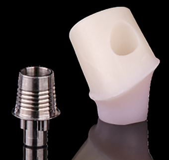
Ceramic abutments versus metal abutments
Abutments are the components of the implant system that connect directly to the implant. Their function is to support the prosthesis, and in doing so they connect the osseointegrated implant to the oral cavity. The mucosa that surrounds the dental abutment has been well studied, and is known to be a stable dimension similar to the biological width that surrounds natural teeth 1,2,3.
The peri-implant mucosa that faces the abutment can be divided into two zones. Firstly, there is the marginal zone which consists of a junctional epithelium approximately two millimetres long, and secondly, a more apical zone containing fibre-rich connective tissue, which is between one to one and a half millimetres high1,4. This soft tissue seal is essential to protect the ossiointegrated implant from the oral environment.
Until recently, dental implant abutments made of titanium have been considered the ultimate treatment for the restoration of an implant, with excellent survival rates from clinical studies5,6,7. However,
their ability to fulfil the criteria of clinical success in the aesthetic zone of subjects with high expectations, a high smile line and a thin gingival biotype is questionable8,9.
Since the introduction of high-strength ceramic abutments in the ı990s, strong interest has grown in them. The main question has been in their ability to perform as successfully and to be as biocompatible as their titanium counterpart10. The benefits of a ceramic abutment are obvious as when they are used, the peri-implant mucosa lacks the greyish discolouration, which can be obvious around metal implant abutments8,11.
While the use and availability of dental implants becomes more widespread, so too does the aesthetic demand. However, many factors are important in the overall success of the dental implant. It is, therefore, important that all factors are taken into consideration when choosing a material for an abutment to ensure optimal outcomes in all definitions of success.
In conjunction with good aesthetics, the abutment material must also be biocompatible and show good clinical performance with high strength and high fracture toughness12. Today, titanium, gold alloy, and zirconium are the main materials used to fabricate implant abutments. They come as prefabricated or customised.
In this paper, we will review the literature relating to metal and ceramic abutments in order to conclude on a preferable material in relation to biological health and clinical performance.
Literature review
The main abutments that have been used and researched in implant dentistry are made of commercially pure titanium, gold alloys, zirconium and aluminium oxide. Clinical success of an abutment has many facets and combines the absence of biological and technical complications.
Technical complications include fracture of the abutment, problems with the abutment screws (loosening or fracture) and abutment/crown combinations.
In the area of biological assessment, soft tissue complications, recession, aesthetic outcome, bone loss and fistula formation are included in the criteria for success of the abutment.
Metal abutments
Gold alloy abutments
In the restoration of an implant, a UCLA-type abutment is often used. It is usually cast in a gold alloy and is designed to engage the implant directly14. A number of studies have carried out research on the biological health of the surrounding mucosa and the clinical success of gold alloy abutments15,3.
Abrahamsson et al concluded from his animal study that gold abutments differed from the controls (titanium abutments) in relation to the soft tissue. He stated that no proper attachment formed around gold abutments, and that soft tissue recession and bone resorption occurred. It was stated that the mucosal seal receded to the implant fixture3. Although this was a well-controlled study, it can be argued that there were only a small number of dogs in the study (five) and the fact that the implants were inserted into recently extracted areas may have influenced the outcome.
Later studies have shown conflicting evidence. In a four-year prospective controlled randomised trial with a split mouth design, it showed that gold alloy abutments preformed as well as titanium abutments in relation to the health of the peri implant tissue15. However, Welander conducted a study, whereby he examined the mucosal barrier at implant abutments of different materials and concluded that soft tissue healing to gold alloy abutments is different to that of titanium and zirconia16. This study suggests that an apical shift of the mucosal seal and the marginal bone occurred around the gold alloy abutments. He showed that at the connective tissue zone, a larger amount of fibroblasts and smaller amounts of collagen were present in contrast to abutments made from titanium and zirconia16. This was a well-controlled animal study, but it is not comparable to a clinical trial.
Titanium abutments
Commercially pure titanium is an alloy of approximately 99 per cent titanium and small amounts of oxygen, iron, carbon and nitrogen. The amount of oxygen determines the grade of the metal and increasing its amount results in an increase in strength and a decrease
in ductility.
Over the years, many studies have highlighted its stable peri-implant mucosal attachments around implant abutments17,7,18,12. It also shows a very high strength with fracture being a rare complication of titanium abutments19. Lang showed that these abutments have a high level of precision at the abutment implant interface20. After years of clinical success, these abutments are used as the control in implant abutment studies due to the good stability of the metal6.
However, today’s ever-increasing high aesthetic demand calls for more than the titanium abutment can deliver. The grey colour of the titanium may be visible through the peri-implant mucosa, especially in patients with thin gingival bio-types as described by Kao21. This may be of aesthetic concern if the implant is located in the anterior section and the patient has a high smile line22,23.
Ceramic abutments
Alumina based ceramics
In the early ı990s it was clear that a ceramic material was needed as an alternative to titanium abutments. The first all-ceramic implant abutments were made of densely sintered, high-purity alumina. Since then, numerous papers have shown a similar response of the peri-implant tissue when the alumina abutment is compared with titanium abutments23. In an animal study carried out by Abrahamsson, results showed that abutments made of alumina ceramic provided a mucosal attachment similar to that of titanium 3.
Unfortunately, there are concerns about its strength. Andersson carried out a five-year prospective study comparing alumina ceramic abutments to titanium abutments. The success rate for alumina at five years was 98.1 per cent compared to 100 per cent for conventional abutments24.
This was due to abutment fracture. Alumina is more brittle, less resistant to tensile forces and it is prone to fatigue. Micro-structural defects in the material may cause cracks in combination with tensile forces25.
Zirconia
Zirconia was introduced into dentistry in the 1990s because of its good mechanical and chemical properties. It is a polymorphic material that occurs in three forms: a cubic structure at its melting point (2680ºC) which transforms into a tetragonal phase below 2370ºC. Below 1170ºC, zirconia is in the monoclinic phase and is accompanied by a 3-5 per cent volume expansion which causes high internal stresses26. This is known as stress induced transformation toughing. The stress associated with expansion acts in opposition to the stress that promotes the propagation of the crack, therefore highly intense tension will be required for the fracture to continue propagating 27.
The mechanical properties of zirconia are the highest ever reported for a dental ceramic. It is therefore an ideal material for the use of an implant abutment because of its high fracture resistance compared to alumina.
The reported use of zirconia since its introduction in 1995 is very promising, with results showing ı00 per cent survival rates19,28,29. A clinical study has yet to report on the fracture of a zirconia abutment34.
Apart from its mechanical properties, zirconia is white in colour, which has obvious benefits for aesthetic reasons. It is also radiopaque and biocompatible.
In an in vivo human study, titanium and zirconium oxide disks were attached to the molar region of ı0 patients30. The surface roughness of both types of disks was the same. After 24 hours the disks were evaluated by scanning electron microscopy for the evaluation of bacterial adhesion. The results showed statistically significant differences between the two materials. The titanium disk resulted in ı9.3 per cent of its surface colonised by bacterial species with a thin layer of cocci covering most of the remaining surface. The zirconia surface was only colonized 12.1 per cent with many areas showing no bacteria or salivary proteins. Rimondini had similar findings in his study31. The probable explanation for this lies in its superficial structure and in its electrical conductivity30. This low colonisation potential, in conjunction with its toughness and aesthetic value, make it a very suitable material
for abutments.
Some studies have shown the biological stability resulting from abutments made from zirconia. Healthy mucosal conditions and stable marginal bone levels were observed at implants with zirconia abutments32,33,28.
Comparing abutment materials
A small number of randomised controlled clinical trials have been carried out comparing abutments of different materials:
Andersson et al 2001
In a study entitled Alumina Ceramic Implant Abutments used for Single Tooth Replacement: A Prospective 1-3 Year Multicenter Study, alumina abutments were compared to titanium abutments. It consisted of two groups: Group A included 69 abutments, 34 test and 35 control, followed for one year. Group B was a subset of group A and consisted of 20 abutments, ı0 test and ı0 control, followed for three years.
Results showing failure rates can be seen in Table One below.

No significant difference was found between test and control abutments for gingival inflammation or bone loss. Aesthetic results were recorded as excellent by both clinician and patient in all cases.
However, the strength of the alumina is the main concern. In this study, 75 per cent of the implants were replacing incisor teeth, and so the alumina abutments were not exposed to the heavier posterior occlusal load. This does not give a good indication of the fracture resistance of the test material. However, it is obvious that the alumina abutments are not as strong as metal.
There was also a mixture between screw retained and cemented crowns which may have skewed the results from a biological point of view. This also resulted in a difference in restorations being all ceramic for cement retained and porcelain on metal for screw retained. The cemented crowns were placed with three different types of cement. The above conditions limit the study results as other factors may have influenced the results. Group B had both test and control abutment with the same restoration and same cement, which gives us a much more stable result, although a much smaller sample size. Here we see no difference in survival between the two abutment types.
Andersson et al 2003
Ceramic Implant Abutments for Short Span FPD’s: A Prospective 5 Year Multicenter Study.
This randomised controlled clinical trial aimed to compare ceramic and metal abutments on implants supporting short-span fixed partial dentures. It gives us more reliable results regarding the strength of the abutment material as the bulk of the FPDs were placed in the posterior region. The results are displayed in Table Two below.

Again, the strength of the ceramic material is questionable. However, in most other fields they preformed as well as the control abutment. Due to the tendency for ceramic materials to undergo fatigue, studies longer that five years need to be carried out to show the true strength of the material.
Vigolo et al 2006
A Four-Year Prospective Study to Assess Peri-implant Hard and Soft Tissue Adjacent to Titanium Versus Gold Alloy Abutments in Cemented Single Implant Crowns.
In this study, 40 implants were placed in 20 patients. Single-tooth bilateral edentulous spaces were the criteria. This study was well controlled, as all test implants, abutments and crowns were the same, as were the controls. In addition, the same technician fabricated the abutments and crowns, and the same prosthodontist restored the implants. The results show no significant differences between the two groups.
The information outline in Table Three differs to some other studies (Abrahamsson 1998) (Welander 2008). However, the above study was very well controlled, apart from the fact that it does not specify how the teeth were lost. If periodontal disease was not a factor, then low pathogenic bacterial counts may have produced this result, where little gingival inflammation and bleeding occurred.
Sailer et al 2009
Randomised controlled clinical trial of customised zirconia and titanium implant abutments for canine and posterior single tooth implant reconstructions: preliminary results at one year of function.
This study was carried out to test the strength of zirconia in posterior and canine regions, and to compare aesthetic results of both abutment types. A total of 22 patients were included with 40 implants. This was a very thorough study and result assessment included many facets. Under the heading of technical complications, this included fracture of the abutment, fracture of veneering porcelain and loss of retention either due to screw loosening or fracture of the screw. Aesthetics was measured by comparing peri-implant mucosal colour to control teeth using a spectrometer. The Jemt papilla index was also used (Jemt 1997). Soft tissue thickness was established using a fine endodontic file. The results are presented in Table Four.
This shows that at one year, zirconia shows the same survival and aesthetic outcome as titanium. This was a very well controlled study, however, only 12 of the original 20 patients from the control group presented for review, and ı9 from the test group attended. It cannot give a true aesthetic result, as the implants replaced posterior and canine teeth, which are not directly in the aesthetic zone and have thicker gingival tissue.
Also, in the inclusion criteria, any patience presenting with evidence of bruxism were excluded. While this helps to control the study, it may falsely represent the population and the strength of the test abutments.

Discussion and conclusion
The literature review reveals a variance in the findings on the performance of titanium and ceramic implant abutments. Titanium abutments have very high clinical success rates, but are not always a suitable abutment choice. The purpose of this review was to determine if ceramic abutments are preferable to metal abutments from both a biological and technical viewpoint.
In arriving at a conclusion, it is important that all the relevant literature is assessed. In a systematic review carried out this year (Sailer 2009) all of the available information on abutments was extracted from the literature and analysed. Table Five summarises the results from 29 clinical studies which included 5,849 abutments.
Inclusion criteria for this review were strict, as from 7,136 titles, only 29 were chosen. However, only 166 ceramic abutments were included in this study. The other 5,683 were metal abutments. Criteria included an observation period over three years, detailed information on abutment type and detailed analysis of the data. This review concluded that no significant differences in the biological or clinical performance of the abutment types occurred and that ceramic abutments can be considered a valid alternative to metal abutments.
From all of the above literature, I conclude that ceramic abutments are preferable to metal abutments, when it comes to an aesthetic region with a thin gingival biotype, high smile line and high expectations.
The performance to date of zirconia strongly suggests that it is a preferable material to metal, showing fewer biological and technical problems and having a similar survival rate to metal, but as yet there are no long-term studies to back up this point of view.
About the author
Dr Sarah Flannery obtained her dental degree from Trinity College, and graduated with honours in 2010. Previously, Sarah studied dental hygiene at Trinity College and qualified with a distinction in 2002. She worked for a number of years as a dental hygienist in a number of specialist practices before returning to further her career in dentistry.
Sarah has been dedicated to continuing her dental education and has attended many courses internationally. Among them are the Dr. Lorenzo Vanini, Master classes in composite dentistry, Lake Como, Italy and Digital Smile Design Courses in Istanbul and Timisoara with the world renowned Dr Christian Coachman and Dr Florin Cofar.
She has also completed a number of courses in areas such as endodontics, paedodontics and orthodontics. She provides a wide range of treatments including six month braces, Digital Smile Design and children’s dentistry.
Sarah is a member of the Irish Dental Association and the International Academy of Aesthetic Dentistry. Areas of interest include cosmetic smile makeovers, minimal prep veneers and adhesive dentistry. Here at Seapoint Sarah’s gentle friendly manor makes her a wonderful choice for any patient who is anxious or nervous.
References
1. Abrahamsson I, Berglundh T, Wennström J, Lindhe J. The peri-implant hard and soft tissues at different implant systems. A comparative study in the dog. Clinical Oral Implants Research 1996 Sep;7(3):212-9.
2. Berglund T, Lindhe J. Dimension of the periimpant mucosa: Biologic width revisited. J Clin Periodontal. 1996;23:971-973.
3. Abrahamsson I, Berglundh T, Glantz PO, Lindhe J.The mucosal attachment at different abutments. An experimental study in dogs..J Clin Periodontol. 1998 Sep;25(9):721-7.
4. Buser D, Weber HP, Donath K, Fiorellini JP, Doppalapudi V, Paquette DW, Williams RC. Comparison of healed tissues adjacent to submerged and non-submerged unloaded titanium dental implants. A histometric study in beagle dogs..Clin Oral Implants Res. 1996 Mar;7(1):11-9.
5. Andersson B, Odman P, Lindvall AM, Lithner B. Single-tooth restorations supported by osseointegrated implants: results and experiences from a prospective study after 2 to 3 years. Int J Oral Maxillofac Implants. 1995 Nov-Dec;10(6):702-11.
6. Henry PJ, Laney WR, Jemt T, Harris D, Krogh PH, Polizzi G, Zarb GA, Herrmann I. Osseointegrated implants for single-tooth replacement: a prospective 5-year multicenter study. Int J Oral Maxillofac Implants. 1996 Jul-Aug;11(4):450-5.
7. Scheller H, Urgell JP, Kultje C, Klineberg I, Goldberg PV, Stevenson-Moore P, Alonso JM, Schaller M, Corria RM, Engquist B, Toreskog S, Kastenbaum F, Smith CR. A 5-year multicenter study on implant-supported single crown restorations. Int J Oral Maxillofac Implants. 1998 Mar-Apr;13(2):212-8.
8. Jung RE Sailer I, Zembic A, , Hämmerle CH, Mattiola A. Single-tooth implant reconstructions: esthetic factors influencing the decision between titanium and zirconia abutments in anterior regions. Eur J Esthet Dent. 2007 Autumn;2(3):296-310.
9. Sailer I, Zembic A, Jung RE, Hämmerle CH, Mattiola A. Single-tooth implant reconstructions: esthetic factors influencing the decision between titanium and zirconia abutments in anterior regions. Eur J Esthet Dent. 2007 Autumn;2(3):296-310.
10. Glauser R, Sailer I, Wohlwend A, Studer S, Schibli M, Schärer P. Experimental zirconia abutments for implant-supported single-tooth restorations in esthetically demanding regions: 4-year results of a prospective clinical study. Int J Prosthodont. 2004 May-Jun;17(3):285-90.
11. Park SE, Da Silva JD, Weber HP, Ishikawa-Nagai S. Optical phenomenon of peri-implant soft tissue. Part I. Spectrophotometric assessment of natural tooth gingiva and peri-implant mucosa. Clin Oral Implants Res. 2007 Oct;18(5):569-74.
12. Sailer I, Philipp A, Zembic A, Pjetursson BE, Hämmerle CH, Zwahlen M. A systematic review of the performance of ceramic and metal implant abutments supporting fixed implant reconstructions. Clin Oral Implants Res. 2009 Sep;20 Suppl 4:4-31.
13. Vigolo P, Majzoub Z, Cordioli G.Measurement of the dimensions and abutment rotational freedom of gold-machined 3i UCLA-type abutments in the as-received condition, after casting with a noble metal alloy and porcelain firing. J Prosthet Dent. 2000 Nov;84(5):548-53.
14. Vigolo P, Givani A, Majzoub Z, Cordioli G.A 4-year prospective study to assess peri-implant hard and soft tissues adjacent to titanium versus gold-alloy abutments in cemented single implant crowns. J Prosthodont. 2006 Jul-Aug;15(4):250-6.
15. Welander M, Abrahamsson I, Berglundh T. The mucosal barrier at implant abutments of different materials. Clin Oral Implants Res. 2008 Jul;19(7):635-41.
16. Lindhe J.Berglundh T, Dimension of the periimplant mucosa. Biological width revisited. J Clin Periodontol. 1996 Oct;23(10):971-3.
17. Abrahamsson I, Zitzmann NU, Berglundh T, Linder E, Wennerberg A, Lindhe J.The mucosal attachment to titanium implants with different surface characteristics: an experimental study in dogs. J Clin Periodontol. 2002 May;29(5):448-55.
18. Andersson B, Taylor A, Lang BR, Scheller H, Schärer P, Sorensen JA, Tarnow D. Alumina ceramic implant abutments used for single-tooth replacement: a prospective 1- to 3-year multicenter study. Int J Prosthodont. 2001 Sep-Oct;14(5):432-8.
19. Lang NP , Lima LA, Fuchs-Wehrle AM, , Hämmerle CH, Liberti E, Pompeu E, Todescan JH. Surface characteristics of implants influence their bone integration after simultaneous placement of implant and GBR membrane. Clin Oral Implants Res. 2003 Dec;14(6):669-79.
20. Kao RT, Fagan MC, Conte GJ. Thick vs. thin gingival biotypes: a key determinant in treatment planning for dental implants.J Calif Dent Assoc. 2008 Mar;36(3):193-8.
21. Yildirim M, Edelhoff D, Hanisch O, Spiekermann H. Ceramic abutments–a new era in achieving optimal esthetics in implant dentistry. Int J Periodontics Restorative Dent. 2000 Feb;20(1):81-91.
22. Henriksson K, Jemt T-30.Evaluation of custom-made procera ceramic abutments for single-implant tooth replacement: a prospective 1-year follow-up study. Int J Prosthodont. 2003 Nov-Dec;16(6):626.
23. Andersson B, Glauser R, Maglione M, Taylor A. Ceramic implant abutments for short-span FPDs: a prospective 5-year multicenter study. Int J Prosthodont. 2003 Nov-Dec;16(6):640-6.
24. Belser UC, Schmid B, Higginbottom F, Buser D. Outcome analysis of implant restorations located in the anterior maxilla: a review of the recent literature. Int J Oral Maxillofac Implants. 2004 19 Suppl:30-42. Review.
25. Piconi C, Maccauro G. Zirconia as a ceramic biomaterial. Biomaterials. 1999 Jan;20(1):1-25. Review.
26. Cavalcanti AN, Foxton RM, Watson TF, Oliveira MT, Giannini M, Marchi GM. Y-TZP ceramics: key concepts for clinical application. Oper Dent. 2009 May-Jun;34(3):344-51. Review.
27. Canullo L .Clinical outcome study of customized zirconia abutments for single-implant restorations. Int J Prosthodont. 2007 Sep-Oct;20(5):489-93.
28. Scarano A, Piattelli M, Caputi S, Favero GA, Piattelli A. Bacterial adhesion on commercially pure titanium and zirconium oxide disks: an in vivo human study. J Periodontol. 2004 Feb;75(2):292-6.
29. Rimondini L, Cerroni L, Carrassi A, Torricelli PBacterial colonization of zirconia ceramic surfaces: an in vitro and in vivo study.Int J Oral Maxillofac Implants. 2002 Nov-Dec;17(6):793-8.
30. Brodbeck U. The ZiReal Post: A new ceramic implant abutment. J Esthet Restor Dent. 2003;15(1):10-23..
31. Kohal RJ, Klaus G. A zirconia implant-crown system: a case report. Int J Periodontics Restorative Dent. 2004 Apr;24(2):147-53.
32. .Sailer I, Zembic A, Jung RE, Hämmerle CH. Randomized-controlled clinical trial of customized zirconia and titanium implant abutments for single-tooth implants in canine and posterior regions: 3-year results. Clin Oral Implants Res. 2009 Aug;20(8):802-8.
33. Jemt T. Regeneration of gingival papilla after single implants. Int. Journal of perio rest dent 1997; (17); 151-157
You must be logged in to post a comment.